A thyroid nodule is a lump in or on the thyroid gland, located in the lower front of the neck. Thyroid nodules are common but often go undiagnosed. They are found in about six percent of women and one to two percent of men, becoming more common with age. While the majority of thyroid nodules are benign, the possibility of cancer must always be considered.
The Features of Thyroid Nodule
Common Occurrence: Found in about 6% of women and 1-2% of men, more common in older individuals.
Usually Asymptomatic: Most patients have no symptoms and nodules are often discovered incidentally.
Lump in the Neck: Can be noticed by patients or during routine physical exams and imaging studies.
Possible Symptoms: Vague pressure sensation or discomfort when swallowing.
Benign Majority: The vast majority of thyroid nodules are non-cancerous
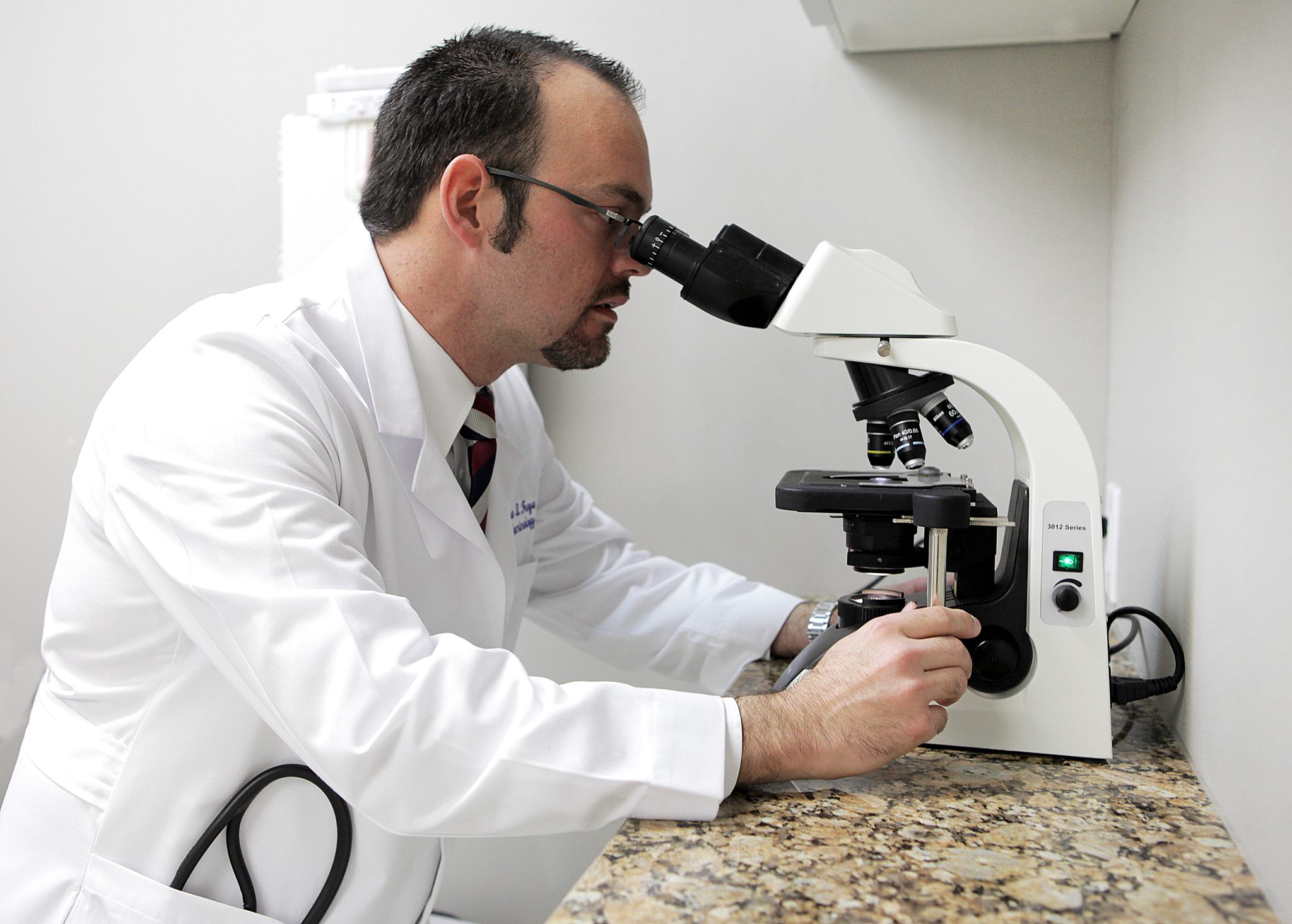
1. Thyroid Needle Biopsy: A thyroid fine needle biopsy involves extracting tissue from the nodule using a thin needle. This minimally invasive procedure provides detailed information, often eliminating the need for surgery. Ultrasound guidance and molecular testing enhance diagnostic accuracy.
2. Thyroid Scan :A thyroid scan uses a radioactive isotope to image the thyroid gland, identifying nodules as hot (hyperfunctioning), warm (normal), or cold (nonfunctioning). Hot nodules rarely indicate cancer. This test helps guide treatment, especially for hyperthyroidism.
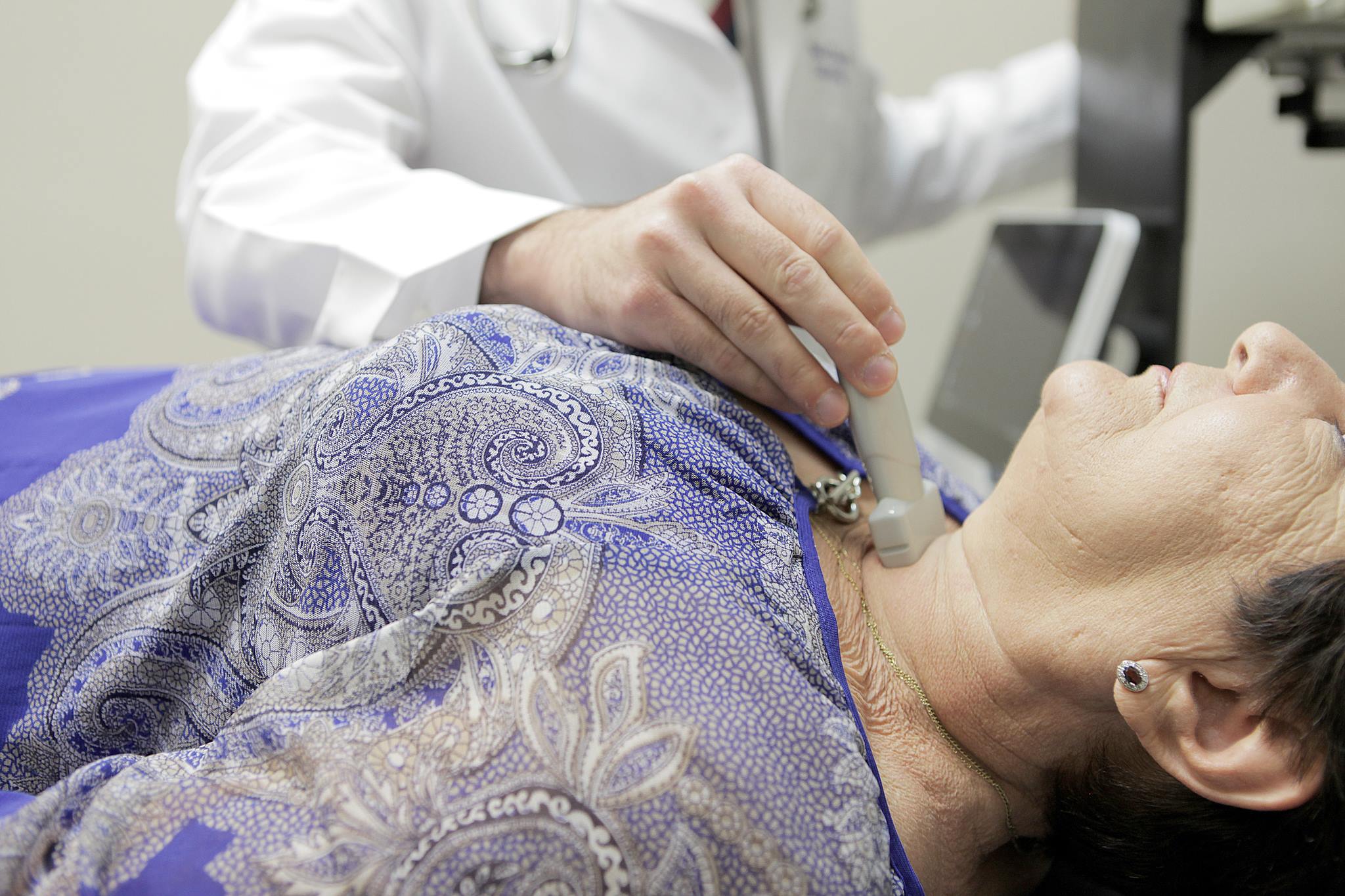
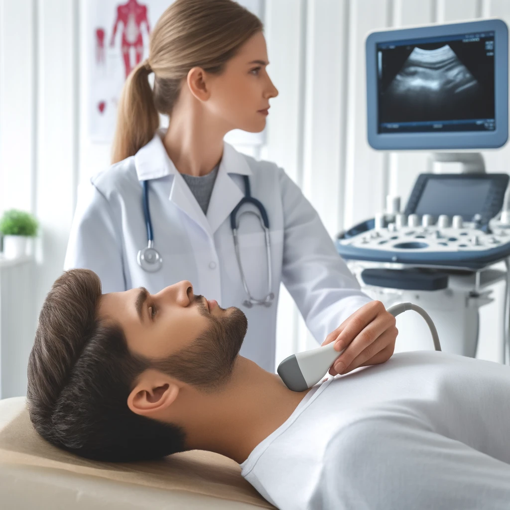
3. Thyroid Ultrasonography: Thyroid ultrasonography uses high-frequency sound waves to create detailed images of the thyroid gland, distinguishing between cystic and solid nodules. It guides fine needle biopsies and monitors nodule size, improving diagnostic accuracy and patient care.
THE TREATMENT
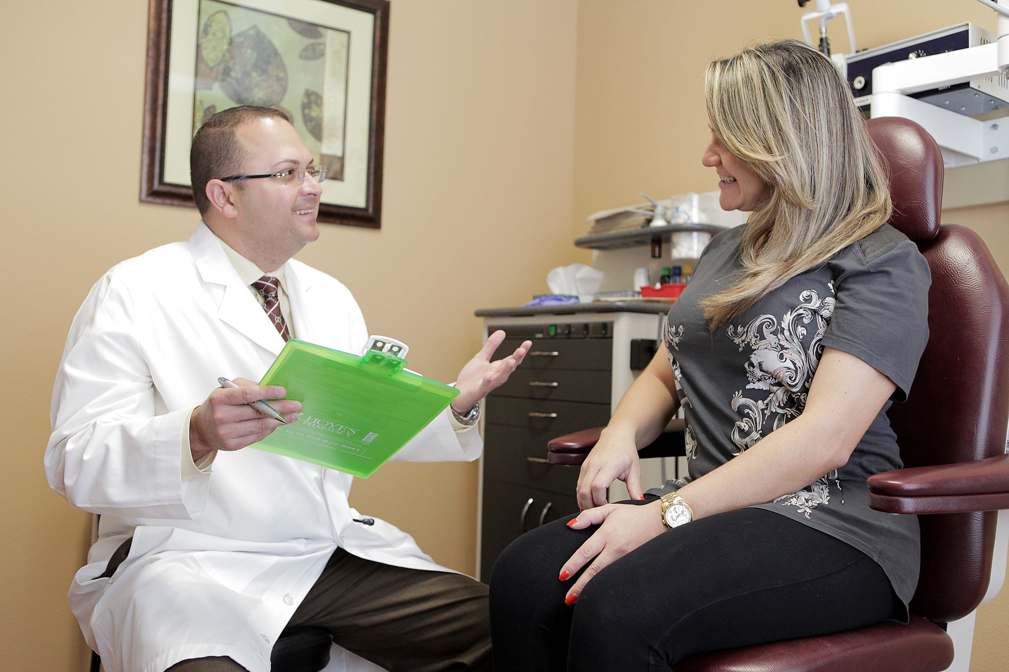
How Are Thyroid Nodules Treated?
Treatment depends on the characteristics of the nodule:
Benign Nodules: Often require no specific treatment and can be monitored.
Levothyroxine Therapy: Sometimes prescribed to prevent nodule growth or reduce the size of cold nodules.
Radioiodine Therapy: Used for hot nodules causing hyperthyroidism.
Surgery: Recommended if cancer is suspected. The goal is to remove cancerous nodules and, if malignancy is confirmed, the entire thyroid gland and any abnormal lymph nodes.
Regular follow-up with an experienced physician is important for monitoring thyroid nodules and ensuring appropriate management.
Need to speak with the Doctor? We’d love to hear from you!
Our Endocrinologists, Dr. Carlo A. Fumero, Sean Amirzadeh, DO, Alberto Garcia Mendez, Lauren Sosdorf, and Pedro Troya, are board certified by the American Board of Internal Medicine and have a wealth of experience treating thyroid conditions. They will work with you to create a personalized treatment plan that meets your unique needs.
As a patient of Dr. Troya’s for several years, I would highly recommend this endocrinologist to anyone. He gives full attention to you and your needs without being pressed for time. His diagnosis and treatments are thorough with up-to-date technology and years of experience. His concern and compassion are genuine with your best interest at heart. After having multiple biopsies performed by Dr. Troya, I am definitely a loyal follower.
Victor Cruz
Successful diabetes treatment, complete care
Read More
I have been going to Dr. Troya for a year. He has treated my Diabetes with great success. He communicates with my other doctors, so he has a complete medical history of all my health issues. We need more caring doctors like Dr. Troya who has the patient’s best interest at heart. I am very thankful for Dr. Pedro Troya.
Michael Hoover
Stabilized diabetes, regained pilot's license.
Read More
I was a private pilot for 34 years before contracting Type 1 diabetes after a severe case of food poisoning in March of 2008. My first endocrinologist refused to assist me in working with the FAA to retain my pilot’s license. I became a patient of Dr. Troya in 2010. Since that time, he has put me on a Continuous Glucose Monitoring regime and helped me stabilize my blood glucose levels to levels that allow me to pass my FAA medical exam. His caring and interest in each patient's personal issues, as well as their medical outcomes, marks him as an outstanding physician.
KC Mullis
Personable, caring, thorough, trusted physician.
Read More
Dr. Carlos Fumero is a very personable and caring physician. He takes the time to listen and is very thorough in his evaluations and explanations. He exercises incredible care and concern. I have been referring family members and friends to Dr. Carlos Fumero for almost two years now. We are all exceptionally grateful to have found such a trusted physician. Thank you, Dr. Carlos Fumero. Your kindness and medical expertise are more appreciated than I could ever express in words.
Previous
Next
Need to speak with the Doctor? We’d love to hear from you!